SRMC is an advanced bioimaging facility at the Life Sciences Institute. It provides access, user training, and technical support for super-resolution microscopy (STED, SMLM, SIM), confocal microscopy (point laser scanning or spinning disk), image processing, 3D reconstruction, and quantitative analysis. We are among the few bioimaging facilities in North America which provide these techniques and tools across the biological length scales. LSI Imaging is a multi-user facility and has been accessed by over 400 researchers from more than 100 research groups, spanning 15 Departments and 7 Faculties across the UBC research ecosystem, from BC Cancer Agency, Djavad Mowafaghian Centre for Brain Health, UBC Hospital, UBC Point Grey campus, SFU, and the industry.
Please see the table for a summary, and click the instruments below the table for specs/more details.

Confocal Microscopes
- The spinning disk confocal system is based on an inverted Zeiss Axiovert 200M microscope equipped with a QuantEM 512SC Photometrics camera
- 443, 473, 515, 561 and 658nm laser lines
- DAPI, FITC, Rhodamine filter cubes for epi fluorescence
- 20X/0.5 EC Plan Neofluar, 63X/1.4 Oil DIC Planapo, 100X/1.45 Oil Plan-Fluor
- Spherical Aberration Correction unit
- Additional high-resolution camera with the capability of taking 1024*1024 images
- Live cell imaging with temperature control
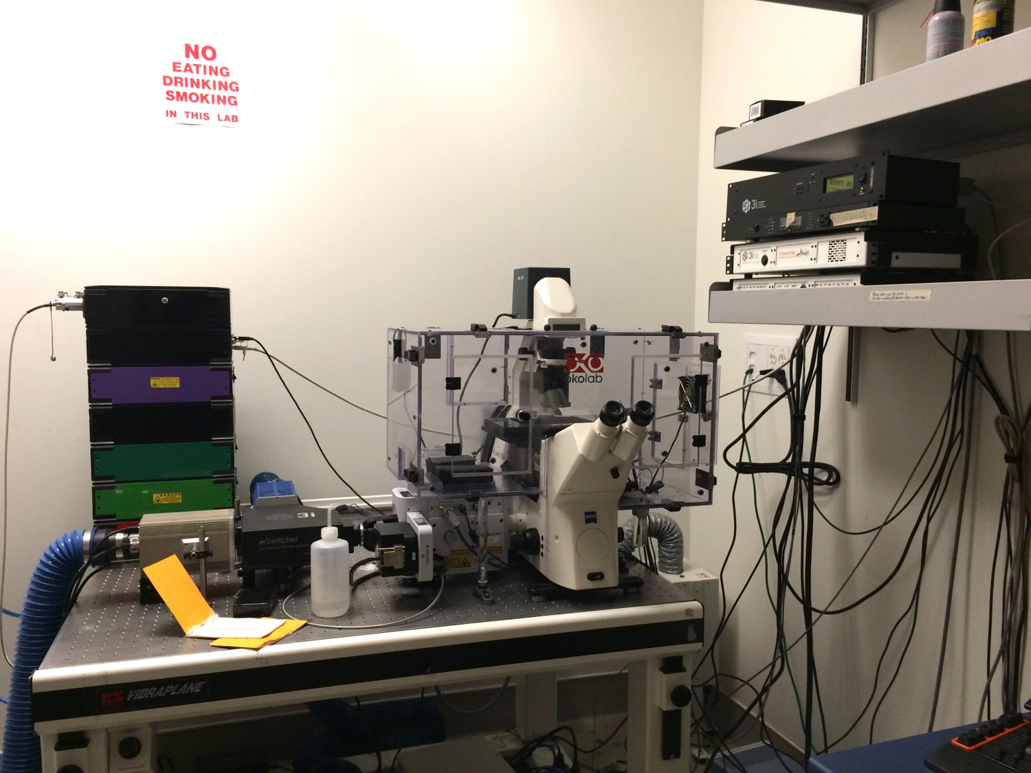
- SIM scanner for simultaneous laser stimulation (with a 405nm laser) and observation (e.g. FRAP)
- Multi-line Argon laser (457, 488, 515nm), 405nm diode laser, 543 and 633nm HeNe lasers
- DAPI, FITC, TRITC filter cubes for epi fluorescence
- Three PMT detectors for fluorescence and one PMT for BF and DIC
- Spectral scanning mode
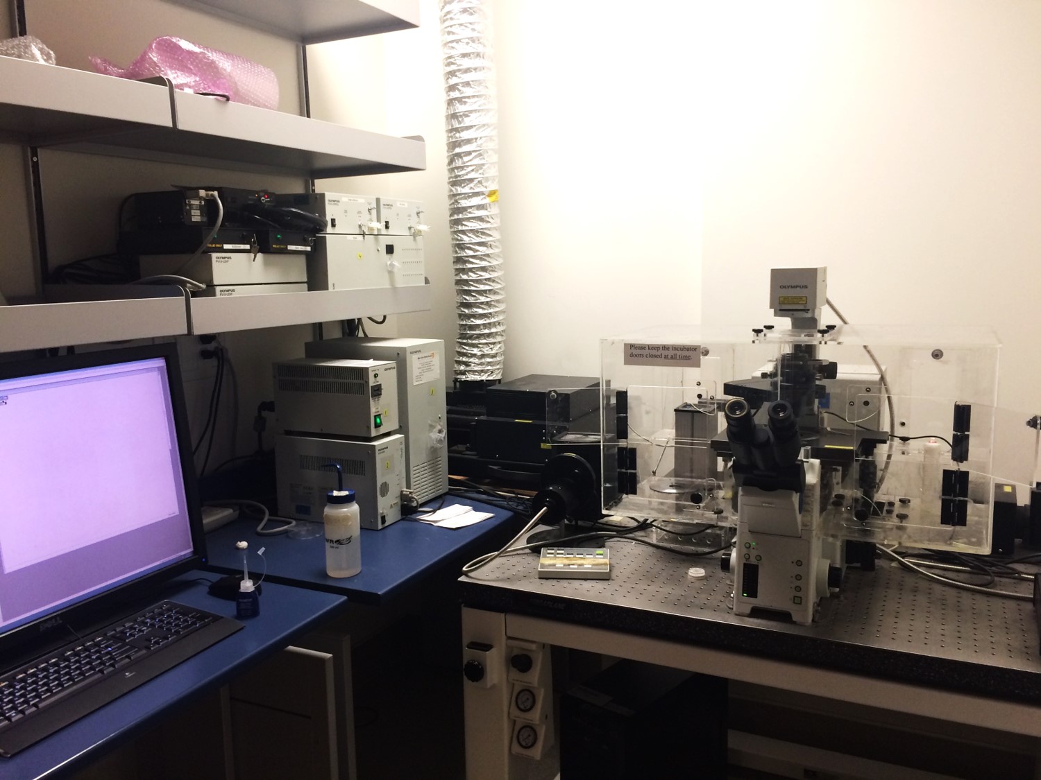
- The confocal system is based on the inverted Olympus IX81 microscope
- UPlanFl 10X/0.30, UPlanApo 20X/0.70, UPlanApo 60X/1.2 Water (corr), UPlan 60X/1.35 Oil and UplanSApo 100X/1.40 Oil
- Live cell imaging with temperature control
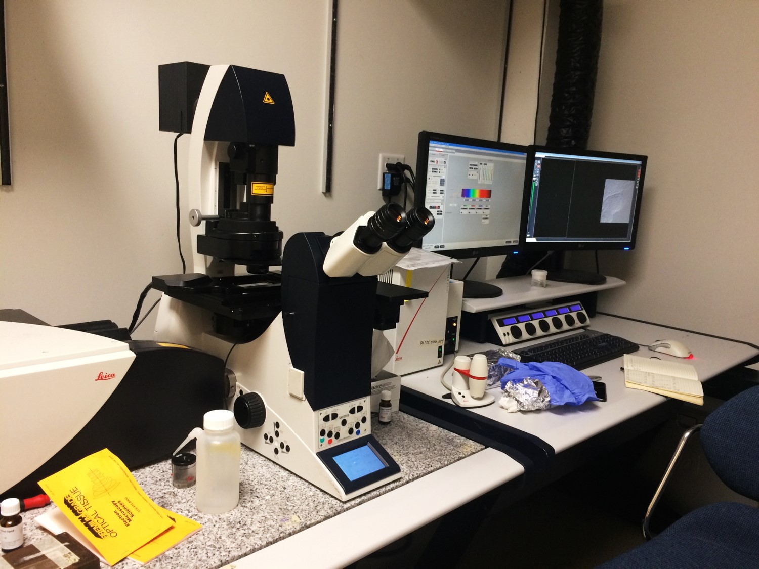
- Based on Leica DMI6000 CS
- Built-in LAS AF for image acquisition and analysis
- Objectives: 10X dry, 20X dry and 63X/1.4 oil
- Motorized XY stage for 3D imaging
- 2.12 mm scanfield diameter with 10x
- Rotation, panning and zooming capability
- Heated enclosed environmental chamber for live imaging
- 4 PMT detectors
- Lasers: 405, 543, 594, 633 and multi-line argon (458, 476, 488, 514)
- FITC and TRITC filter cubes for epi fluorescence
- STELLARIS 5 LIAchroic is a true confocal point scanning system with LIAchroic beam splitters. It includes a highly sensitive, prism-based spectral detection design with computer controlled adjustable bandwidth for all fluorescence channels.
- Lasers: 405, 448, 488, 561, 638 nm
- Objectives: HC PL FLUOTAR 10x/0.30, HC PL APO 20x/0.75 CS2, HC PL APO 63x/1.40 OIL CS2, 25x water IMM long working distance
- LAS X 3D
- Live cell imaging with temperature and CO2 controls
- LAS X Lightning Expert
- LAS X Dye Finder
- 4 Power HyD S detector
- Inverted research microscope with touch screen based on the DMi8 series
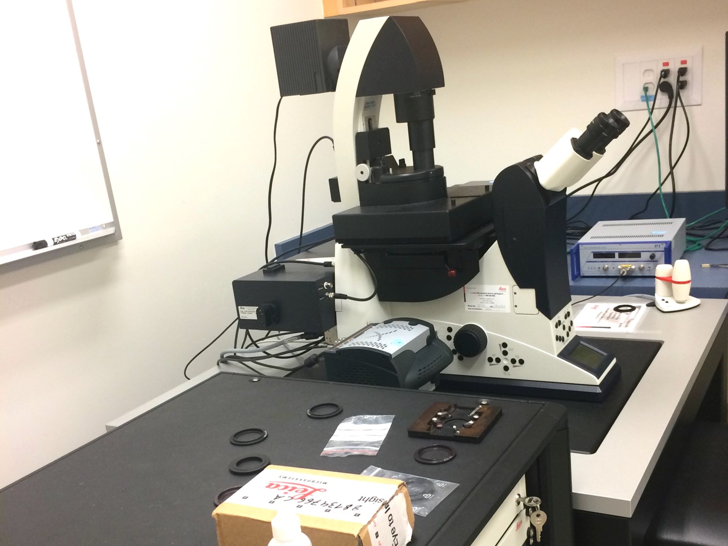
- Leica SP5 is based on Leica DMI6000 CS
- High-speed scanning
- DIC, DIC-Pol, Bright field
- Built-in LAS AF for image acquiring and analysis
- Objectives: 20X dry, 40X oil, 60X dry, 60X water and 63X/1.4 oil
- 4 PMT detectors
- High-precision z-glavo and motorized XY stage for 3D and high throughput imaging
- Lasers: 405, 488, 561, 633 nm
- DAPI, FITC, TRITC filter cubes for epi fluorescence
Super-Resolution Microscopes
- Single molecule localization-based super-resolution microscopy system
- 488, 532 and 642 nm laser lines with 500 mW laser power
- 405 nm backpumping laser line; 160x objective lens
- High speed, high sensitive EMCCD camera
- The lateral resolution of up to 20 nm, the axial resolution of up to 50 nm
- TIRF capacity with all three laser lines
- Super-resolution system based on a laser scanning confocal with donut-shaped depletion lasers.
- White light laser (WLL: full spectra of 470-670 nm excitation lines) and 405 nm individual laser line.
- Spectral scanner and high-speed resonant scanner.
- 10x air, 20x air, 60x water, HC PL APO 86x/1,20 W motCORR STED white and HC PL APO 100x/1,40 OIL STED WHITE objective lenses.
- PMT and Leica HyD high-sensitivity detector.
- High-precision z-glavo stage.
- 592 nm, 660 nm, and 775 nm STED depletion laser lines which allow multiple channels super-resolution imaging.
- Time-gating confocal and STED (gSTED) with XY resolution down to 30nm.
- Live-cell imaging (temperature).
- AFC for focus control in long-term live-cell imaging.
- DAPI, FITC, TRITC filter cubes for epi fluorescence.
- Leica LAS X Life Science Microscope Software
- FLIM and tauSTED
- FRET

Please contact the Core facility if you are interested in dSTORM

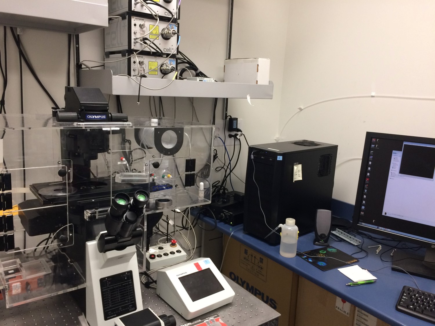
- Olympus cell^TIRF system based on Olympus IX83 automated inverted microscope.
- Cell^TIRF illuminator enable motorized control of TIRF angles, precise adjustment of incident angle and software calculation of penetration depth for different wavelengths.
- Built on MetaMorph software platform.
- Three diode-pumped solid-state laser lines - 405nm, 491 nm and 561 nm with simultaneous excitation of two desired lines. Integrated Point FRAP optics for 405nm laser line. Filter sets for epifluorescence - DAPI, GFP and RFP
- Platform for live cell imaging, full enclosure for temperature control and humidity incubator; no CO2 [can be added on request]
- Three TIRF objectives – planApo 60X, NA 1.45; UApoN 100X, NA 1.49, WD 0.09mm, Correction collar; and UApoN 150X, NA 1.45, WD 0.07mm, Correction collar
- Two high performance Photometrics Evolve 512*512 pixels EMCCD cameras equipped with DC2 emission splitting system, for fast acquisition of two spectrally distinct images simultaneously. Acquisition speeds of 33 frames/sec in 512*512 format, 66 frames/sec in 256*256 format and up to 100 frames/sec in smaller formats.
- Ideal for high speed single molecule imaging and membrane research
High Content Fluorescence Analysis
Features:
- High Resolution CCD Camera
- Standard 5X, 10X, 20X and 40X objectives
- Standard 6-Color Capability
- High Efficiency Mercury-Xenon Illumination
- Carl Zeiss AxioVert optical Bench: • Apoptome • Twister robot arm
BioApplications include automated analysis of a wide range of biological events.
- Target Activation
- Compartmental Analysis
- Morphology Explorer
- Acquire Only
- Micronucleus
- Cell Motility
- Cell Spreading
- GPCR signal
- Spot Detector
- Cytotoxicity
- CytoCellMemTrans
- Neuronal Profiling
- Colocalization
- Cell Health Profiling
- Molecular Translocation
- Nuc Trans
- Tube Formation
For more information regarding Cellomics equipment and modules, please visit their website at www.cellomics.com
To become a user or to ask/report something with the system, contact Dr. Michel Roberge by email or at 604-822-2304.
Click here to view the rules for use. Only registered users for this system can book online. New users are encouraged to familiarize themselves with the rules as inappropriate access and usage may result in loss of booking privileges.
 Cost: $15/hr
Image Analysis Workstation
- Image-Pro Plus V6.3 with SharpStack 3D deconvolution package.
- Slidebook V4.2.
- ImageJ.
- Huygens Professional deconvolution software
- ImageJ 2 (Fiji) with ThunderSTORM & QuickPALM plugins
- Imaris-3/4D Image Visualization and Analysis Software
- Adobe package for figure preparation
- Nikon Elements (NIS-Elements)
Additional Imaging Systems at the LSI
Contact indicated individual if you like to use a system
- Gen II Multidimensional Imager with Prior automated stage.
- Roper Scientific Coolsnap HQ camera.
- CFP, CY5, DAPI, FITC, FRET 436/535, FRET CFP 436/470, TRED, TRITC, YFP, ZSensorBright and ZSensorDim.
- Sutter Instrument Corp. Xenon illumination.
- 4X – 60X dry objectives.
To become a user or to ask/report something with the system, please contact Dr Francois Jean by mail or at 604-822 0256.
- Bruker Biospec 70/30 USR Preclinical MRI system.
- Compatible specimen size: dependent on the size of gradient coils chosen. For typical applications, gradient coils of 12 cm or 20 cm inner diameter are used.
- Typical resolution achievable in a standard anatomical scan: 100 microns in-plane, 500 microns through-plane.
- 7.05 Tesla magnetic field at isocentre of cylindrical magnet bore.
- Scan gating and physiological monitoring system (ECG, respiration, temperature).
- Multi-nuclear NMR capability: 1H, 31P, 19F, 13C.
- 4 Receiver channels and parallel imaging capabilities.
The UBC MRI Research Centre operates a state of the art 7 Tesla MRI scanner in the Life Sciences Centre. This instrument is capable of performing imaging experiments and NMR spectroscopy on in vivo specimens. We are a user-supported facility which aims to help academic and industrial scientists meet their research goals through the use of MRI technology, particular for the study of animal modles of human disease and disorder.
Booking the system: The instrument is supported by full-time staff who will run the MRI experiments according to the investigator’s needs and study design. Please visit the User’s Guide to the 7T Facility for more information.
Example applications:
- Body fat imaging and non-imaging measurements of lean-to-fat mass ratio
- Imaging of cells in vivo with MRI-visible tracers (e.g. iron oxide labels)
- Morphological/anatomical imaging
- NMR spectroscopy in brain, heart and tumour, for measurements of metabolite concentrations
- Water diffusion tensor imaging
- Perfusion MRI to measure hemodynamics and vascular parameters
Widefield Microscopes
- With a Leica DMi8 stand with a motorized stage for large sample imaging (tile scan)
- 5x, 10x, 40x DRY objective, 60x oil and 100x oil objectives
- Very sensitive Leica K8 camera;
- Leica K3C color camera for H&E staining;
- Wide field imaging for up to 8 channels;
- THUNDER processing on the fly, small volume and large volume features for thick samples.
- DIC, BF.
- PTI DeltaRAM V monochromator.
- Filters are CFP/YFP/dsRed, Fura-2, GFP, Fura-2/dsRed, CFP/YFP, EGFP/dsRed.
- The system is based on an inverted Olympus IX71 microscope equipped with a Photometrics Cascade 512B camera.
This system is not an LSI Imaging system, however the owner Dr Robert Nabi is willing to share the system with other users. If you like to use this system, please contact Robert Nabi by mail or at 604-822-7000 to set up training and/or to reserve a time slot.
- Automated Zeiss 200m microscope with motorized Z-stage for wide field fluorescence microscopy with 3D deconvolution and deblurring.
- Ultra-fast Sutter Lambda DG4 xenon excitation source.
- Nomarski Differential Interference Contrast Microscopy at 100x and 40x.
- Roper CoolSnapHQ2 cooled CCD camera.
- Emission filter wheel-based Forster Resonance Energy Transfer (FRET).
- imaging and a wide variety of filters for multiple FRET pairs.
- Laser-based Fluorescence Lifetime Imaging (FLIM) in the frequency domain (lasers at 440 and 532 nm).
- Laser-based Total Internal Reflection Fluorescence Microscopy (TIRFM) (lasers at 440 and 532 nm).
- Individual filter cubes for DAPI, FITC, Cy3, TxRed, and Cy7 allowing multicolour labelling with minimal bleedthrough.
- Zeiss Objectives: a) 100x Oil (1.45 NA), b) 40x Oil, c) 40x Air Long Working Distance, d) 20x Air, e)10x Air, f)5x Air, g)1.25x Air.
- Temperature controls and solution perifusion for live cell imaging.
- Eppendorf semi-automatic single-cell microinjection and flash photolysis for caged compounds.
- Slidebook software and offline analysis computer.
- To be installed – motorized x/y-stage.
This system is not an LSI Imaging system, however the owner Dr Jim Johnson is willing to share the system with other users. If you like to use this system, please contact Dr Johnson by mail or at 604-822-7187 to set up training and/or to reserve a time slot.
System 1: Widefield microscope for single and dual wavelength fluorescence measurements in live cells.
Zeiss Axiovert 10 epifluorescence microscope (inverted) with 40x long-working distance and 63x oil immersion objectives. Interfaced with Atto Biosciences intensified CCD camera (8 bit, 512 x 480 pixels), camera controller, filter changer, high speed shutter and RatioVision software. Temperature-controlled superfusion chamber (accepts 18 or 22 mm round glass coverslips) and associated pumps, etc. Stand alone analysis software (PC only).
This system is designed for [ion]i measurements in live cells using single excitation (e.g. fluo-3 for [Ca2+]i) or dual excitation (e.g. fura-2 for [Ca2+]i, BCECF for pHi, SBFI for [Na+]i, PBFI for [K+]i) probes. Although very sensitive, the camera on this system has limited spatial resolution (reliable recordings can be obtained only from structures and/or cells >5 µm diam.).
System 2: Widefield microscope for single wavelength, dual wavelength and dual emission fluorescence measurements in live cells.
Zeiss Axiovert 135 epifluorescence microscope (inverted) with 40x long-working distance and 63x oil immersion objectives. Interfaced with two Atto Biosciences intensified CCD cameras (each 8 bit, 512 x 480 pixels), camera controller, filter changer, high speed shutter and RatioVision software. Temperature-controlled superfusion chamber (accepts 18 or 22 mm round glass coverslips) and associated pumps, etc. Stand alone analysis software (PC only).
This system can perform the same [ion]i measurements (and has the same limited spatial resolution) as System #1. The addition of a second camera and filters allows the use of dual emission (e.g. indo for [Ca2+], SNARF for pH) as well as dual excitation probes. A particular advantage of this system is the ability to measure the intracellular concentrations of more than one ionic species simultaneously (e.g. Sheldon et al., J. Neurosci. 24, 11057-11069, 2004). Automated routines are available for the simultaneous measurement of [Na+]i or [K+]i + pHi; [Ca2+]i + pHi; [Cl-]i + pHi; [Na+]i or [K+]i + [Ca2+]i; [Ca2+]i + [cAMP]i; and [Ca2+]i + mitochondrial membrane potential (e.g. Fernandes et al., J. Neurosci. 27, 13614-13623, 2007). Other routines are easily developed and electrophysiological recordings can be performed simultaneously with dual [ion]i measurements (e.g. Kelly & Church, J. Neurophysiol. 96, 2342-2353, 2006).
System 3: Widefield microscope for single and dual wavelength fluorescence measurements in live cells.
Zeiss Axioskop 2 FSPlus microscope (upright) with 20x, 40x, 63x and 100x water-dipping objectives. Interfaced with a QImaging Retiga EXi cooled CCD camera (12 bit, 1360 x 1036 pixels), Sutter DG5 high speed filter changer and shutter, and Intelligent Imaging Innovations software (Slidebook). Temperature-controlled superfusion chamber (accepts 15 mm round glass coverslips) and associated pumps, etc. Stand alone analysis software (PC only).
This system can perform the same [ion]i measurements as System #1, with the added advantage of high spatial resolution. DIC images can be captured during the course of ratiometric [ion]i measurements, making this system particularly useful for examining changes in [ion]i at the leading edge of migrating cells or neuronal growth cones.